Your brain is not a static organ; it’s constantly evolving. Throughout your life, learning and new experiences continuously reshape it. This remarkable ability of the brain to reorganize itself by forming new neural connections in response to learning, experience, or environmental changes is called neuroplasticity, also known as brain plasticity or neural plasticity.
Most learning processes within the brain involve intricate rewiring. This means creating and strengthening connections between neurons, the fundamental cells in your brain responsible for learning and information processing. Imagine neurons as tiny messengers, and learning as building stronger pathways for these messengers to communicate more effectively.
While the majority of neurons in most brain regions are present from birth and remain with you throughout life, research indicates that neurogenesis, the growth of new neurons, continues in at least one small but significant area: the hippocampus. The hippocampus plays a vital role in learning and memory. However, the precise function of these newly generated neurons in learning is still an active area of research.
Neuroplasticity is fundamental to all forms of learning. Neuroscience research in this field has extensively studied how the brain recovers from injury or damage. Interestingly, many of the principles that govern brain recovery also apply to how the brain adapts and changes with learning throughout your entire life. This means that the brain’s capacity for change isn’t limited to healing; it’s also the foundation of our ability to learn and grow.
The Dynamic Relationship Between Learning, Memory, and Brain Change
To understand how learning affects your brain, let’s consider a fundamental principle: learning and memory are intrinsically linked. You cannot learn without storing information in some form of memory. This stored information becomes accessible for future use, whether it’s recalling knowledge or improving skills. Neuroscience reveals that memories are encoded through physical changes in the brain. While the exact nature of these changes is still under investigation, it’s clear that your brain undergoes physical modifications whenever learning occurs. Therefore, every experience and learning opportunity throughout your life physically shapes and molds your brain.
There’s a common misconception that the brain reaches full development in early childhood, implying that further change is minimal. This myth suggests that brain development in childhood and adolescence is primarily predetermined by biology, culminating in a fixed state in adulthood.
However, the reality is far more nuanced. Brain development is a dynamic interplay between your genetic blueprint and your unique learning experiences. Biological factors do not equate to predetermined outcomes. Your brain is continuously sculpted by your experiences. It is never static but remains adaptable, changing in response to learning throughout your entire lifespan.
A Historical Perspective: Unveiling Brain Structure and Debunking Myths
In the late 19th century, Santiago Ramón y Cajal, a pioneering biologist, proposed the neuron theory. This theory posited that the brain is composed of discrete yet interconnected cells, much like the cells that constitute the rest of the body. Initially, his contemporaries ridiculed this idea, believing the brain to be fundamentally different. However, subsequent research validated Cajal’s theories, earning him the Nobel Prize in 1906.
The brain is indeed made up of cells called neurons, densely connected through synapses. The majority of neurons reside in the cortex, or grey matter, the brain’s outer layer, only a few millimeters thick. The characteristic folds of the brain maximize the surface area of the grey matter, allowing a vast number of neurons to be packed within the skull.
Figure 1. Exquisitely detailed hand-drawings by Ramón y Cajal (1899) illustrate the brain’s fundamental cells (neurons) and their extensive interconnections (synapses) within the surface layer (grey matter), revealing the brain’s intricate structure.
Despite his groundbreaking contributions, Cajal made one significant error. He claimed that “In adult centers the nerve paths are something fixed, ended, immutable. Everything may die, nothing may be regenerated.” While he was partially correct that damaged neurons are not repaired or regenerated, this led to the myth that the brain becomes fixed after early adulthood, only declining with age.
Contrary to this myth, the brain is never “fixed, ended, and immutable.” It continuously changes with learning throughout life, primarily by modifying the connections, or wiring, between neurons.
A compelling example of brain plasticity is seen in London taxi drivers. They demonstrate an exceptional ability to navigate the complex city without maps, a skill reflected in their larger-than-average hippocampi, the brain region associated with spatial memory. This showcases how acquiring and utilizing knowledge physically alters brain structure.
Synaptogenesis: The Power of Brain Connections
The immense computational power of the brain stems from its vast network of interconnections between neurons, facilitated by synapses. While the brain houses approximately 86 billion neurons, each neuron can connect to thousands of others, resulting in an estimated 150 trillion synapses.
The number of neurons remains relatively constant throughout life and is not significantly altered by learning or experience. Instead, the connections between neurons, the synapses, are constantly changing. These synaptic changes are the primary mechanism for learning and memory in the brain. These changes involve two key processes:
- Synaptogenesis: The formation of new synapses, creating new pathways for neural communication.
- Long-Term Potentiation (LTP): The strengthening of existing synapses, enhancing the efficiency of communication along specific neural pathways.
Much of our understanding of synaptogenesis comes from animal studies. Donald Hebb, a prominent neuroscientist in the 1940s, observed that rats raised in stimulating environments, resembling their natural habitats, outperformed those raised in standard laboratory cages on cognitive tasks. Providing rats with ample physical, social, and sensory stimulation led to more extensive interconnections between their neurons and a greater number of synapses.
While direct evidence in humans is limited, studies on children from severely deprived Romanian orphanages in the 1980s revealed persistent cognitive, language, and social development delays, highlighting the detrimental effects of severe deprivation on brain development. Interestingly, research on Albert Einstein’s brain suggests he had a more interconnected brain, with denser neuron connections between the left and right hemispheres, possibly facilitating more efficient brain communication.
Hebb also described Hebbian learning (1949), a crucial principle of brain plasticity summarized as “neurons that fire together, wire together.” Simply put, when two or more neurons are repeatedly activated simultaneously by a thought, action, or environmental event, the synapse between them strengthens. This strengthens the association between those neurons. In the future, if one of these neurons is activated, it becomes more likely to trigger a response in the connected neurons, recalling and reinforcing the associated memory or skill.
Thus, a significant portion of learning in the brain involves modifying synaptic connections, particularly reinforcing frequently used pathways or circuits of interconnected neurons.
Neurogenesis: The Ongoing Birth of New Neurons
While most of the brain’s neurons are present from birth, one specific area, the hippocampus, continues to generate new neurons throughout life through neurogenesis. The hippocampus is well-established as a critical region for memory and learning.
It’s only in recent decades that research has confirmed adult neurogenesis in the human hippocampus. A groundbreaking study using carbon-dating techniques to determine the age of individual hippocampal cells estimated that approximately 700 new neurons are added to each hippocampus (left and right) daily. By age 60, about one-third of the neurons in the hippocampus are newly generated after birth.
This discovery has generated considerable excitement and is at the forefront of neuroscience research. However, much remains unknown about the precise role of these new neurons in learning and memory.
We know that the hippocampus is essential for forming new memories. Damage to the hippocampus results in severe amnesia, impairing the ability to form new memories after the damage occurred. The hippocampus also plays a crucial role in spatial navigation, our ability to remember and navigate familiar environments, a discovery recognized with the 2014 Nobel Prize.
Factors like exercise, a healthy diet, stress reduction, and learning itself are known to promote neurogenesis. While numerous books, websites, and products claim to “boost your brain” by enhancing neurogenesis, the direct benefits of specifically targeting neurogenesis for cognition, memory, or learning are still under investigation. Currently, there’s insufficient evidence to definitively claim that these methods will directly “boost your brain.”
The key takeaway is that the hippocampus continuously generates new neurons throughout life and is vital for learning and memory. However, whether enhancing neurogenesis directly translates to increased intelligence or cognitive ability is a question for future research.
Neuroplasticity in Action: Real-World Examples
The brain’s capacity for reorganization through neuroplasticity is remarkable. It rewires, alters, and strengthens connections and pathways that are frequently used. As Hebb’s principle describes, frequently used pathways of interconnected neurons strengthen their connections and become “wired together.”
Much neuroplasticity research focuses on how the brain recovers from damage or injury. For example, the brain regions controlling body movement and touch sensation have body maps called homunculi. Damage to the motor cortex, for instance, due to stroke, can cause weakness in corresponding body parts.
While damaged neurons do not regenerate, and new neurons don’t grow in these regions, individuals can regain movement control. Rehabilitation and repeated practice of movements enable undamaged brain areas to remap their connections and take over functions from the damaged areas. This is the basis of physiotherapy for movement rehabilitation, building new pathways as the brain relearns movement control through new connections.
The principles of creating and strengthening connections also apply to typical learning, not just recovery from brain damage.
For example, a study on string instrument players revealed they have a larger sensory area in the brain dedicated to touch sensation in their left hand compared to their right hand or non-musicians. Extensive practice with left-hand finger movements on strings molds the brain, creating and strengthening connections, allocating more brain area to the left hand’s sensory input.
In another study, young adults learning to juggle for three months showed an increase in grey matter size in a brain area important for perceiving moving objects. After they stopped juggling, this area returned to its original size within three months. This size change wasn’t due to new neuron growth, as neurogenesis doesn’t occur in this brain region. Subsequent research suggested that changes in connections within the grey matter were responsible for this plasticity during juggling skill acquisition.
These examples demonstrate the brain’s immense adaptability and capacity for change through altering and strengthening connections based on experience and use. Frequent use of specific brain pathways strengthens those pathways. This is considered a primary mechanism for brain learning – adapting and changing connections based on experience.
The Enigma of Einstein’s Brain: A Case Study in Neural Connectivity
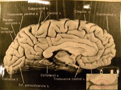
When Albert Einstein passed away in 1955, his brain was removed during autopsy, photographed, dissected, and preserved. The subsequent history of these preserved parts is complex, with many sections still unaccounted for. However, in 2010, a collection of original autopsy photographs resurfaced and were acquired by the National Museum of Health and Medicine, Washington, DC, USA.
Figure 2. A photograph of Albert Einstein’s brain, captured during the 1955 autopsy. These photographs, lost for years, were recovered in 2010 and have been used in recent research to investigate the unique structural features of his brain.
Recent research has utilized these original photographs to analyze the density of neuron connections between the left and right hemispheres of Einstein’s brain, specifically examining the corpus callosum, a structure facilitating interhemispheric communication. Researchers compared Einstein’s corpus callosum, based on the photographs, to MRI scans of individuals of the same age as Einstein at his death (76 years old) and individuals of Einstein’s age during his “miracle year” of 1905 (26 years old), when he published groundbreaking scientific papers.
The findings revealed that Einstein’s corpus callosum was thicker in most regions compared to both older and younger comparison groups. This suggests Einstein had more extensive neural connections between his brain’s hemispheres. The researchers concluded that Einstein’s exceptional intellectual abilities might be linked to enhanced communication and coordination between the two hemispheres of his brain.
Educational Implications: Embracing Brain Plasticity in Learning
These fundamental neuroscience principles highlight the brain’s remarkable capacity for change through learning. While bridging the gap between these basic principles and the complexities of education, knowledge acquisition, and skill development (like reading and mathematics) is still a significant undertaking, these core concepts of neurons, synapses, and dynamic neural connections are at the heart of all brain-based learning.
Several immediate educational implications emerge from these principles:
- The brain is always ready to learn. Biological predispositions do not limit learning potential. The brain continuously adapts and changes with learning and experience throughout life; it never becomes fixed or unchangeable. Therefore, “smartness” is not solely determined by biology but significantly shaped by brain connectivity resulting from learning.
- Learning is an inherent brain function. The brain learns automatically through everyday experiences, tasks, problem-solving, and habit formation. Frequently used brain pathways strengthen their connections, reinforcing memories and improving skills. While deliberate practice enhances learning, the fundamental principles of connection change and strengthening apply whenever neural pathways are engaged.
- Effective learning emphasizes connections. Deliberate learning should focus on establishing and strengthening connections between related concepts rather than rote memorization of isolated facts. Connecting new information to existing knowledge networks enhances understanding and retention.
References
- Cajal, S. Estructura de los centros nerviosos de las aves. 1-10 (Jiménez y Molina, 1888).
- Colucci-D’Amato, L., Bonavita, V. & di Porzio, U. The end of the central dogma of neurobiology: stem cells and neurogenesis in adult CNS. Neurological Sciences 27, 266-270 (2006).
- Maguire, E., Woollett, K. & Spiers, H. London taxi drivers and bus drivers: A structural MRI and neuropsychological analysis. Hippocampus 16, 1091-1101 (2006).
- Cajal, S. Comparative study of the sensory areas of the human cortex. (Clark University, 2013).
- Hebb, D. Committee on Graduate and Professional Training. American Psychologist 2, 206-206 (1947).
- Blakemore, S. & Frith, U. Learning Brain. 32-32 (Wiley, 2005).
- Men, W. et al. The corpus callosum of Albert Einstein‘s brain: another clue to his high intelligence? Brain 137, e268-e268 (2014).
- Cajal, S. Comparative study of the sensory areas of the human cortex. (Clark University, 1899).
- Spalding, K. et al. Dynamics of hippocampal neurogenesis in adult humans. Cell 153, 1219-1227 (2013).
- Lieberwirth, C., Pan, Y., Liu, Y., Zhang, Z. & Wang, Z. Hippocampal adult neurogenesis: Its regulation and potential role in spatial learning and memory. Brain Research 1644, 127-140 (2016).
- Dr. Ananya Mandal, M. Hippocampus Functions. News-Medical.net (2019). at
- Elbert, T., Pantev, C., Wienbruch, C., Rockstroh, B. & Taub, E. Increased cortical representation of the fingers of the left hand in string players. Science 270, 305-307 (1995).
- Draganski, B. et al. Changes in grey matter induced by training. Nature 427, 311-312 (2004).