The human skull is a complex structure comprised of 22 bones that provide protection for the brain and support for the face. Learning these bones can seem daunting, but with effective strategies, it can become an engaging and manageable task. This article provides a comprehensive guide on How To Learn The Skull Bones, covering various memorization techniques and helpful resources.
Understanding the Skull’s Structure
Before diving into memorization, it’s crucial to understand the skull’s basic organization. The skull is divided into two main parts:
- Cranium: Encloses and protects the brain. It consists of eight bones: frontal, two parietal, two temporal, occipital, sphenoid, and ethmoid.
- Facial Skeleton: Forms the framework of the face. It comprises 14 bones, including the mandible (lower jaw), maxilla (upper jaw), zygomatic bones (cheekbones), and nasal bones.
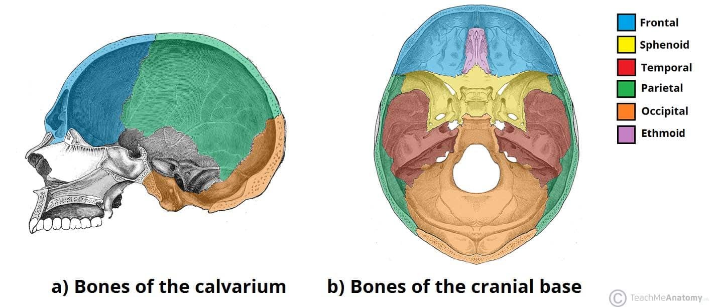
Fig 1: The cranium can be further divided into the cranial roof (calvarium) and cranial base.
Effective Memorization Techniques
Several methods can help you effectively learn the skull bones:
- Visual Aids: Utilize anatomical diagrams, 3D models, and even real skulls (if accessible) to visualize the bones’ shapes, sizes, and relationships.
- Repetition and Review: Consistent repetition is key. Regularly review the material using flashcards, quizzes, and practice labeling diagrams.
- Mnemonics: Create acronyms, rhymes, or stories to associate the bone names with memorable phrases or images. For example, “Paris Eats Tasty Oranges” to remember the parietal, ethmoid, temporal, and occipital bones.
- Chunking: Break down the skull into smaller regions (e.g., cranial base, facial bones) and learn the bones within each region before moving on.
- Active Recall: Test yourself frequently without looking at your notes. This helps solidify your memory and identify areas needing further review.
- Spaced Repetition: Review material at increasing intervals to optimize long-term retention.
Fig 2: The pterion, a clinically significant area where four skull bones meet, is a weak point susceptible to fractures.
Utilizing Resources
Numerous resources can aid in your learning process:
- Anatomy Textbooks: Provide detailed information on skull anatomy and often include helpful illustrations.
- Online Anatomy Atlases: Offer interactive 3D models and labeling exercises.
- Anatomy Apps: Mobile apps provide convenient access to learning materials and quizzes on the go.
- Videos and Tutorials: Visual learners can benefit from watching videos that explain skull anatomy and demonstrate memorization techniques.
Fig 3: The facial skeleton provides structure for the face and houses important sensory organs.
Clinical Relevance and Fractures
Understanding the skull bones is essential in clinical settings, particularly when dealing with fractures.
- Cranial Fractures: Can result from blunt force trauma and may damage underlying structures like the middle meningeal artery.
- Facial Fractures: Common injuries often caused by falls or impacts. Nasal bone fractures are the most frequent type. Maxillary, mandibular, and zygomatic arch fractures are also common and can be classified using specific systems like the Le Fort classification.
Fig 4: Nasal fractures are the most common type of facial fracture.
Sutures and Fontanelles
Skull bones are connected by fibrous joints called sutures. These sutures are immovable in adults but are flexible in infants, allowing for skull growth. Fontanelles are soft spots in an infant’s skull where the sutures haven’t fully fused.
Fig 5: Fontanelles allow for brain growth and skull deformation during childbirth.
Conclusion
Learning the skull bones requires dedicated effort but is achievable with a combination of understanding, visualization, and memorization techniques. Utilizing various resources and focusing on clinical relevance can enhance the learning process and make it more engaging. Consistent practice and review are essential for long-term retention.